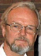
On February 17, Heuser speaks on “Imaging activities inside living cells by quick-freezing and electron microscopy.” The presentation is part of a session called “Frontiers in biological imaging.” The session runs from 8:30 a.m. to 11:30 a.m.
John Heuser, professor of cell biology and biophysics, pioneered a technique for imaging cells and molecules in the electron microscope (EM) that he calls the “quick-freeze deep-etch” procedure. The process has allowed him and his colleagues to take highly detailed pictures of rapid events that occur inside our bodies, including the communication that occurs between nerve cells, the uptake and secretion of materials into and out of cells, the mechanism of entry of dangerous viruses into cells, and the rapid movements of cells ranging from contracting muscle cells to swimming sperm.

“Most of the processes that we image occur in a cellular environment that is watery and transparent, which is completely unsuitable for electron microscopy,” Heuser explains. “We have to do something to rigidify the cell and give it contrast before it can be seen with an EM.”
Heuser wanted to improve on classical approaches to solving this problem, which had changed little since the 19th century and involved embalming samples and staining them with heavy metals.
“That sticks everything together into a confusing jumble, far removed from the beautiful natural order we expect to see in the living cell,” he explains.
Heuser compares his “quick-freezing” approach to using a stroboscopic flash to freeze the action in a photograph. Like the strobe, quick-freezing takes about one ten-thousandth of a second, so it can effectively stop any biological event in midstream.
To enable EM scanning of quick-frozen samples, Heuser developed methods for exposing their interiors and for bringing out contrasts in the structures within by coating them with ultrathin films of metallic platinum that mold snugly to their surface contours.
“Our ‘deep-etch replicas’ therefore faithfully reflect the structure of the original quick-frozen tissue but eliminate the need to keep the tissue frozen while it is imaged in the EM,” Heuser explains. “In fact, we discard the tissue before viewing the replica in the EM.”
The environment inside most EMs makes preservation of frozen tissue samples difficult or impossible, Heuser notes.
“Today, only a handful the most expensive and most powerful electron microscopes in the world are capable of viewing frozen biological samples, and these can still only handle the tiniest of cells — bacteria and protozoa, not the nerves, muscles and glands that we really need to study in current medical research,” he says.
To help other researchers circumvent the great expense and formidable limitations of such ‘frozen-cell electron microscopes,’ Heuser and colleagues have worked in recent years to make the equipment and procedures he has developed for his quick-freeze deep-etch procedure available to researchers around the world. He has recently helped set up facilities similar to his laboratory at the University of Siena in Italy, Yale University and the National Institutes of Health.
Washington University School of Medicine’s full-time and volunteer faculty physicians also are the medical staff of Barnes-Jewish and St. Louis Children’s hospitals. The School of Medicine is one of the leading medical research, teaching and patient care institutions in the nation, currently ranked third in the nation by U.S. News & World Report. Through its affiliations with Barnes-Jewish and St. Louis Children’s hospitals, the School of Medicine is linked to BJC HealthCare.