A computerized atlas has brought unprecedented sensitivity to the search for brain structure changes in a genetic condition known as Williams syndrome, revealing 33 abnormalities in the folding of the brain’s surface. The disorder, which occurs in 1 in every 20,000 births, impairs visual and spatial skills but preserves musical ability and sociability.
The findings, published in The Journal of Neuroscience this week, suggest the same technique may produce insights into more common brain development disorders such as autism, according to the researcher who developed the brain atlas at Washington University School of Medicine in St. Louis.
“We already have a study of autism well along in the pipeline with colleagues at the University of California-Davis,” says lead author David Van Essen, Ph.D., the Edison Professor of Neurobiology and head of the Department of Anatomy and Neurobiology. “We think that study will also highlight several previously unrecognized abnormalities in the folding of the cerebral cortex.”
A more detailed inventory of changes in brain folding and its connections to changes in cognitive function should enable researchers to better understand the origins of developmental brain disorders and begin devising new approaches to treat them.
Van Essen announced the creation of the brain atlas, known as the Population-Average, Landmark and Surface-based (PALS) Atlas, in summer 2005, when it was made available online. PALS is the first atlas that accurately portrays the complex folds of the cerebral cortex not just from a single individual but from a group of individuals. This is important because the folding of cerebral cortex varies dramatically from one person to the next, similar to the variability of human fingerprints.
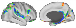
Van Essen and colleagues from Washington University, Stanford University and Cedars-Sinai Medical Center in Los Angeles used data from brain scans of 16 individuals with Williams syndrome for the study. For the analysis, they aligned or “registered” each individual brain to the PALS Atlas. This allowed them to identify 33 changes in the folds of the cerebral cortex, the surface layer of the brain credited with many higher cognitive functions.
“We already knew that there are structural abnormalities in the brains of individuals with Williams syndrome,” notes Van Essen. “What is interesting and new is that we found a plethora of changes discernible on a background of normal variability in folding patterns, and the fact that the changes are strikingly symmetric.”
Scientists found 16 changes on the left side of the brain and identified 16 changes in corresponding regions on the right side of the brain. Another abnormality was present only on the right side of the brain.
Any one person with Williams syndrome would be unlikely to have all 33 changes, Van Essen explains, and the degree of change present can also vary. For example, they found that the olfactory sulcus, a groove or furrow-like structure just above the olfactory tract, tends to be shallower on average in those with Williams syndrome.
Genetic and environmental differences, reactions to injury, and inherited disorders can all change the topography of the brain in minor and major ways. According to Van Essen, the new study shows that PALS can help scientists look beyond such individual variations to quantify brain structure trends in ways that may provide important insights.
Williams syndrome results from deletion of genetic material on a region of chromosome 7, but the size of this deletion varies across individuals. The new inventory of structural changes may one day enable scientists to more closely associate genetic alterations with the development of different brain structures, or allow them to more precisely link alterations in specific structures to changes in cognitive functions.
Although interventions to alter brain development in Williams syndrome are likely still a long way off, identifying the connections between genetic changes, alterations in brain structure and changes in brain function may help clinicians and teachers develop customized approaches to education that allow children with Williams syndrome to take full advantage of their unique capabilities, according to Van Essen.
Van Essen D, Dierker D, Snyder A, Raichle ME, Reiss A, Korenberg J. Symmetry of cortical folding abnormalities in Williams syndrome revealed by surface-based analyses. The Journal of Neuroscience, May 17, 2006.
Funding from the National Institutes of Health, the National Institute of Mental Health, the National Institute for Biomedical Imaging and Engineering, and the National Science Foundation supported this research.
Washington University School of Medicine’s full-time and volunteer faculty physicians also are the medical staff of Barnes-Jewish and St. Louis Children’s hospitals. The School of Medicine is one of the leading medical research, teaching and patient care institutions in the nation, currently ranked fourth in the nation by U.S. News & World Report. Through its affiliations with Barnes-Jewish and St. Louis Children’s hospitals, the School of Medicine is linked to BJC HealthCare.