It’s almost time for the movie to start. As you hurry from the lobby into the darkened theater, you may have to stop as the transition from light into darkness renders you temporarily blind. Cells in the eye’s retina must adapt before you can begin to distinguish heads from backs of chairs. As the cells adjust, sight will be restored enough to avoid tripping over a chair or sitting in a stranger’s lap.
Scientists have long known that these cells, called photoreceptors, are involved in this adaptive process, but a study from investigators at Washington University School of Medicine in St. Louis and Boston University School of Medicine has uncovered a new pathway in the retina that allows photoreceptor cells to adapt following changes in light exposure.
The discovery could help scientists better understand human diseases that affect the retina, including age-related macular degeneration, the leading cause of blindness in Americans over the age of 50. That’s because the process of adapting to darkness involves the same cells that are affected in macular degeneration and other blinding retinal diseases.
The findings are reported online in the journal Nature Neuroscience and will be the cover story in the March print edition.
The retina’s two main light-sensing cells are the rods and cones. Both use similar mechanisms to convert light into vision, but they function differently. Rods are highly sensitive and work well in dim light, but they can quickly saturate with light and stop responding. They don’t sense color either, which is why we rarely see colors in dim light.
Cones, on the other hand, allow us to see colors and can adapt quickly to stark changes in light intensity. The research team focused on cone cells, studying their ability to continue functioning in very bright light and to adapt quickly when that light is shut off.
“Rods can take up to an hour to adapt to darkness,” says principal investigator Vladimir J. Kefalov, Ph.D., assistant professor of ophthalmology and visual sciences. “Cones, by contrast, adapt in three to five minutes.”
Scientists have known for years that light-sensing molecules bind together to make up visual pigments. Those pigments are destroyed when they absorb light and must be rebuilt, or recycled, for the cone cells to continue sensing light. In order to be recycled, key components of pigments called chromophores leave the retina and travel to the eye’s retinal pigment epithelium where the chromophore is restored and returned to the retina.
“If the chromophores cannot be recycled, cone cells gradually run out of visual pigment and can’t detect light,” Kefalov says.
But the process of traveling to and from the pigment epithelium takes too long to explain how cones quickly adapt to darkness following exposure to bright light, so Kevalov’s team went looking for a second, supplementary pathway.
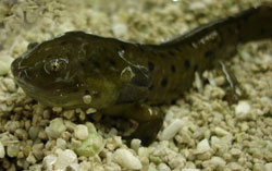
Working in salamander eyes, the research team removed the pigment epithelium layer so that pigment molecules could not be recycled using the known pathway. When the scientists exposed the retina to bright light and then to darkness, the cones continued to function, even without the pigment epithelium. That meant the pigment molecules were recycled in spite of the fact that they could not travel to the pigment epithelium.
“So it was clear that a second pathway is being used by the cone cells,” Kefalov says.
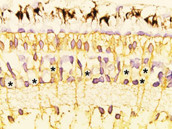
But where? Biochemical evidence had suggested cells called Müller cells might be involved. Like glial cells in the brain that support and interact with neurons, Müller cells in the retina support and interact with photoreceptors. The researchers treated salamander retinas with a chemical that destroyed the Müller cells.
They repeated the experiment exposing the retina to bright light, followed by darkness. “And by blocking the function of Müller cells, we prevented the recycling of chromophores. The cones ran out of photopigment and could not adapt to dark,” Kefalov says.
The group then conducted the same series of experiments in the mouse retina, with the same results, suggesting the second pathway involving Müller cells also is important in the mammalian eye. If it is active in the human eye, Kefalov believes it may be possible one day to manipulate this pathway to improve vision when the other one involving pigment epithelium has been interrupted by injury or disease.
One such disease is age-related macular degeneration, where cone cells begin to malfunction over time. Because the disease and the pathway Kefalov’s team identified both involve cones, he says it may be possible someday to target that pathway, rev up its activity and supplement or rescue the function of cones.
Before that happens, he says it will be important to figure out exactly how the Müller cells are interacting with photoreceptors. Although these studies confirmed the existence of the second photoreceptor pathway, they didn’t reveal how it works. Kefalov’s laboratory has recently received a five-year, $1.9 million National Eye Institute grant to study how this newly identified visual pathway functions in the retina.
Wang JS, Estevez ME, Cornwall MC Kefalov VJ. Intra-retinal visual cycle required for rapid and complete cone dark adaptation. Nature Neuroscience, published online Feb. 1, 2009; http://www.nature.com/neuro/journal/v12/n3/abs/nn.2258.html
This study was supported by the National Eye Institute of the National Institutes of Health, Research to Prevent Blindness and by the Karl Kirchgessner Foundation.