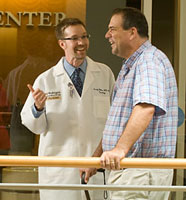
The wasted muscles and slurred speech of advancing ALS (amyotrophic lateral sclerosis), the tremors and movement difficulties of Parkinson’s disease, the slow melting of minds beset by Alzheimer’s disease — such bundled symptoms are so striking that most people assume that the neurological afflictions they signal are unrelated. Not so, says Mark P. Goldberg, MD, director of the Hope Center for Neurological Disorders.

“Patients with brain or spinal disease should of course seek treatment from clinicians specializing in their disorder, but researching specific diseases is not the fast path to a cure. Research shows that diseases of the brain, spinal cord and nerves have common threads that can only be discovered on a fundamental level. When we understand these shared disease pathways, we can begin to find cures. That is the work of the Hope Center.”
Center without walls
Given the complexity of the central nervous system itself — a dynamic, multiscaled and sensitive array of profoundly integrated structures and processes — the Hope Center mandates research in-put from many disciplines. It includes geneticists, molecular, cell and developmental biologists, pathologists, engineers, biochemists and clinician-scientists such as neurosurgeons, pediatricians, internists, psychiatrists, radiologists and anesthesiologists.
In every sense “a center without walls,” the Hope Center facilitates research by 70 scientists on Washington University’s medical and Danforth campuses. The Hope Center’s administrative core is in the Biotechnology Building, and its Program on Protein Folding and Neurodegeneration will be situated in the new BJC Institute of Health at Washington University, located at the heart of the medical campus.
The Hope Center supports innovative research programs through seed grants that effectively leverage traditional grants and contracts as well as private funding. Basic research and scholarly publication, while thriving, are only the beginning of the process. The Hope Center’s guiding principle is translational research: supporting the creation of new knowledge about the brain and nervous system and then enabling its rapid translation into cures, new treatments and diagnostic tools for clinicians and patients.
“When scientists with different perspectives and training focus on a problem together, the translational process accelerates,” says Goldberg, who is professor of neurology, anatomy and neurobiology, and biomedical engineering.
Hopeful response to the times
The Hope Center developed at an intersection of influences. One was Hope Happens, a foundation the late Christopher Wells Hobler established in 2002 to quickly find a cure for ALS patients like himself. Another was the Center for the Study of Nervous System Injury, established in 1991 by former Washington University faculty member Dennis W. Choi, MD, PhD.
At the same time, Goldberg and colleagues like David M. Holtzman, MD, the Andrew B. and Gretchen P. Jones Professor and head of the Department of Neurology, had been impressed with how much progress resulted from collaborative work across disciplines — an approach that Washington University had encouraged for decades. They wanted to implement such a sys-tem to solve the staggeringly difficult questions about brain disease.
“In 2004, Hope Happens approached us about working together,” Goldberg recalls. “It was good timing for both groups. We had a plan in place, and it was just what they were looking for. Neurological science was advancing so rapidly that it was time to begin thinking hard about moving quickly to treatments.”
Organizing the science
To that end, the Hope Center set out to explore neurological diseases’ common mechanisms and effects on brain cells at the level of genes and molecules. Two basic research themes developed. The first, the Hope Center Program on Protein Misfolding and Neurodegeneration, is based on the idea that in neurodegenerative diseases, proteins somehow fold incorrectly after they are formed and then create problems as they aggregate. The program will be one of five Interdisci-plinary Research Centers (IRCs) formed under BioMed 21, the university’s translational research initiative. Led by Holtzman and Alison Goate, PhD, the Samuel and Mae S. Ludwig Professor of Genetics in Psychiatry, the IRC involves 25 researchers whose teams will occupy five laboratories in the new BJC Institute for Health building — the hub for BioMed 21 — when the School of Medicine and Barnes-Jewish Hospital open its doors in January 2010.
Among numerous researchers who have made notable discoveries is Timothy M. Miller, MD, PhD, assistant professor of neurology and director of the Hope Center’s Christopher Hobler Laboratory. Miller’s innovative therapy for ALS that targets the mechanism of protein misfolding has advanced to human trials.
The second major research thrust at the Hope Center is the Program in Axon Injury and Repair. Investigators are seeking to understand how neuronal axons degenerate — with the new realization that when an axon is damaged, the fiber itself triggers a new pathway of active degeneration that could be interrupted with an entirely new kind of treatment. Jeffrey D. Milbrandt, MD, PhD, the David Clayson Professor of Neurology, found that a particular molecule arrests the process and then described the pathway; therapies have since been licensed for clinical development. And in April 2009 — in another example among many — Aaron DiAntonio, MD, PhD, associate professor of developmental biology, discovered a complementary second pathway leading to axon degeneration, suggesting treatments with powerful potential.
“We’re getting so close to truly understanding neurodegenerative disorders and are mak-ing headway with new therapies,” says Holtzman, who credits the Hope Center’s infrastructure for its success in both research and funding. He chairs a steering committee of senior scientists (Goate, Goldberg, Milbrandt and Eugene M. Johnson Jr., PhD, professor of neurology and of molecular biology and pharmacology) to evaluate progress, with oversight from the Hope Center’s executive committee. Matthew J. Stowe, JD, administrative director, coordinates the overall team effort.
Eliminating barriers
Still another way the Hope Center ensures progress is by knocking down conventional barriers, creating smoother, faster pathways to translation. In addition to putting the right minds together to solve complicated problems — such as matching basic scientists with clinicians — administrators have provided core facilities for animal models, amyloid-beta microdialysis, neuroimaging and transgenic and viral vectors. New facilities, equipment and instrumentation — most recently, the medical school’s first atomic force microscope — are added, funding permitting, in response to investigators’ needs. And a new collaboration with the Office of Technology Management recently has been implemented to help scientists disclose and patent inventions and to ready their ideas for biotechnology or drug company licensing.
“In one sense, it’s good that our researchers are distributed across the campuses,” says Goldberg. “They can work near their home departments without changing affiliations and neighbors. The only downside is that while we gather regularly, we don’t interact every day. Bumping into people in a hallway can be at the heart of science. Finding new ways to bring scientists together — now, there is a challenge!”
Tracking gene mutation to treat its effects
Robert H. Baloh, MD, PhD, Assistant Professor of Neurology
Hope Center Program in Mitochondria and Bioenergetics
Robert H. Baloh, MD, PhD, deciphers the molecular mechanics of neurodegenerative disease. Two of his research focuses are amyotrophic lateral sclerosis (ALS), also known as Lou Gehrig’s disease, and Charcot-Marie-Tooth disease (CMT). As neurons die in the motor pathways of an ALS patient’s brain and spinal cord, muscles atrophy. The patient becomes profoundly weak, unable to speak or swallow, and eventually dies from respiratory failure three to five years after diagnosis. In contrast, though CMT involves degeneration of similar motor pathways, it progresses slowly over a patient’s lifetime, typically causing foot drop, foot deformities, falls and hand weakness as peripheral nerves deteriorate.
Baloh’s lab is investigating a gene known as TDP-43, a key regulator of messenger RNA splicing, which edits protein-building instructions from DNA to allow proper protein assembly. Abnormalities can radically alter cellular function.
During collaborative research published in Annals of Neurology in February 2008, Baloh and Hope Center colleagues, including Nigel J. Cairns, PhD, research associate professor of neurology and of pathology and immunology, and Alison Goate, PhD, professor of neurology and of genetics, linked a mutation in TDP-43 to an inherited form of ALS and are now creating a mouse model for the disease. They will then compare animal models, cultured neurons and cell lines to existing models of ALS based on mutations in the SOD1 gene.
“Our expectation,” Baloh says, “is that understanding how disease mutations in TDP-43 and SOD1 cause neurodegeneration will allow us to identify effective treatments.”
In pursuit of brain and spinal cord repair
Valeria Cavalli, PhD, Assistant Professor of Neurobiology
Hope Center Program in Axon Injury and Neurodegeneration
Valeria Cavalli, PhD, a specialist in neuronal cell biology and axon injury, is engaged with one of medicine’s great challenges: to reverse paralysis and restore nerve function when the central nervous system (CNS) has been severely damaged by stroke, spinal-cord injury or disease. Whereas peripheral nerves, like most of the body’s tissues, can usually repair themselves, damaged neuronal axons in the CNS that normally deliver essential electrochemical payloads cannot spontaneously regenerate. When axon damage interrupts transport — of mitochondria, cytoskeletal polymers, neurotransmitters — the delicate conduit degenerates and mental and physical functioning follow suit.
Cavalli wants to know how neurons in the peripheral nervous system manage to regenerate and learn what is different about them and their response to injury. She aims to determine whether they somehow sense injury and their counterparts in the CNS do not—or whether neurons in each system “sense” but only one responds in a particular way.
After dissecting the molecular events from the point of peripheral nerve damage to self-repair, Cavalli intends to determine which steps in CNS response sequences are deficient and devise ways to assist, perhaps by bypassing or stimulating the appropriate mechanism.
“My findings will apply to many neurodegenerative diseases, in which axon damage is due not to traumatic injury but to the effects of protein aggregation or impaired function, which then cause degeneration.”
Tracing molecular missteps
Rohit V. Pappu, PhD, Associate Professor of Biomedical Engineering
Hope Center Program on Protein Folding and Neurodegeneration
Part of the work of biophysicist Rohit V. Pappu, PhD, concentrates on how proteins clump together. This research is relevant to Huntington’s disease — which affects balance, speech and muscle strength, and typically causes death within 20 years — as well as eight other inherited neurodegenerative diseases. All are technically termed polyglutamine expansion disorders, and that means that the affected person produces a protein with an abnormal stretch containing many units of the amino acid glutamine.
In research that answered a longstanding question about why proteins rich in polyglutamine should aggregate, Pappu and coworkers showed that despite the purported “water-loving” nature of glutamine, polyglutamine molecules behave like readily aggregating “greasy” molecules. These findings were made using novel fluorescence measurements coupled with polymer theory and computer simulations.
Further work showed that this aggregation depends on the length of the polyglutamine stretch. “We showed that the inability of individual polyglutamine molecules to fold into well-defined three dimensional structures promotes aggregation,” Pappu says. “By aggregating, the polyglutamine molecules interact to achieve structures that individual polyglutamine molecules cannot achieve on their own.”
Pappu is also part of a project led by Jin-Moo Lee, MD, PhD, associate professor of neurology, and Carl Frieden, PhD, professor of biochemistry and molecular biophysics. The study showed that cells take up small amounts of amyloid beta (Abeta) — peptides that make up ex-tracellular plaques in brains of people with Alzheimer’s disease. Using neuroblastoma cells — malignant but easy-to-work-with cells representative of neurons — the researchers administered Abeta to cells in small, physiologically relevant amounts. They found that the cells took up Abeta and packed it into lysosomes, specialized acidic pouches within cells that digest unwanted proteins. However, the acidic conditions and confined space within lysosomes provide conditions that are conducive to Abeta aggregation, whereby “nature’s protection appears to end up becoming a problem.”
This story appeared in the Summer 2009 issue of Outlook magazine.