The National Institutes of Health awarded a biomedical engineer at Washington University in St. Louis a four-year, $1.7 million grant to attempt to develop a new way to image airflow in lungs. If such research proves successful, it someday could make diagnoses of lung disease considerably more expeditious, easy and cost-effective.
“There is a great clinical need for being able to image ventilation, however, there’s really no easy way to image ventilation in vivo right now. So the purpose of this project is to develop a new technique,” said Mark Anastasio, professor of biomedical engineering at the School of Engineering & Applied Science.
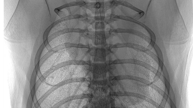
Physicians currently use CT scans and MRI to image airflow — or ventilation — in the lungs. Both methods require the patient to inhale a contrast agent, a procedure that can cause complications. Additionally, such methods require expensive equipment that limit their widespread use.
Anastasio will use sophisticated modeling and machine learning to develop new technology that would enable airflow imaging with a single X-ray image. The method would take less time and not require contrast inhalation beforehand. It potentially would be a less expensive, more efficient way to get a better look at how air circulates through the lungs.
A simple-to-implement imaging method that could provide spatially- and temporally-resolved information regarding ventilation would be of great value to those studying basic pulmonary physiology and the onset and progression of a large range of respiratory diseases in pre-clinical model. It would also facilitate drug discovery and efficacy studies aimed at mitigating respiratory pathology.
“At the end of the day, we want to produce an enhanced X-ray image with values that tell us how much air there is in different parts of the lung. We believe this rapid imaging approach will allow earlier diagnosis of a whole host of lung conditions, including COPD and even lung cancer,” Anastasio said.
Anastasio is working on the project with two colleagues at Washington University School of Medicine: Buck Rogers, a professor of radiation oncology and of radiology, and Steven Brody, MD, the Dorothy R. and Hubert C. Moog Professor of Pulmonary Medicine and a professor of radiology. Together, the researchers plan to evaluate the technology in mice.
“The partnership with the Medical School is critical,” Anastasio said. “Having access to collaborators there with very specific skills and sets of expertise is vital for technology developers like me. It allows our teams to create and evaluate the technology in a very meaningful and systematic way.”
Their work is supported by NIH grant R01EB023045.
The School of Engineering & Applied Science at Washington University in St. Louis focuses intellectual efforts through a new convergence paradigm and builds on strengths, particularly as applied to medicine and health, energy and environment, entrepreneurship and security. With 88 tenured/tenure-track and 28 additional full-time faculty, 1,200 undergraduate students, 1,200 graduate students and 20,000 alumni, we are working to leverage our partnerships with academic and industry partners — across disciplines and across the world — to contribute to solving the greatest global challenges of the 21st century.
Comments and respectful dialogue are encouraged, but content will be moderated. Please, no personal attacks, obscenity or profanity, selling of commercial products, or endorsements of political candidates or positions. We reserve the right to remove any inappropriate comments. We also cannot address individual medical concerns or provide medical advice in this forum.