3D printed, transplantable organs may sound like science fiction, but thanks to advances in polymer chemistry at Washington University in St. Louis, they could become a reality.

Stimuli-responsive hydrogels represent a broad class of soft materials that change their mechanical properties when certain external triggers are applied. Last year, researchers from the lab of Jonathan Barnes, assistant professor of chemistry in Arts & Sciences, created a new kind of artificial molecular muscle from a polymer that changes color and contracts when exposed to blue light. Similar materials promise a wide range of applications, particularly in medicine.
In a new study published recently in the journal ACS Applied Materials & Interfaces, the Barnes lab presented a new kind of responsive polymer that can activate very precise locations in the gel, instead of all of it.
Faheem Amir, lead author on the paper and a postdoctoral researcher in the Barnes lab, reported success in several areas. “The process resulted in significant increases in the soft material’s stiffness, tensile strength, and percent elongation before breaking, all of which could easily be reversed via oxidation and swelling in water,” he said. The hydrogels also allowed precise spatial resolution and control over where activations took place, which the team illustrated by photopatterning an American flag design.
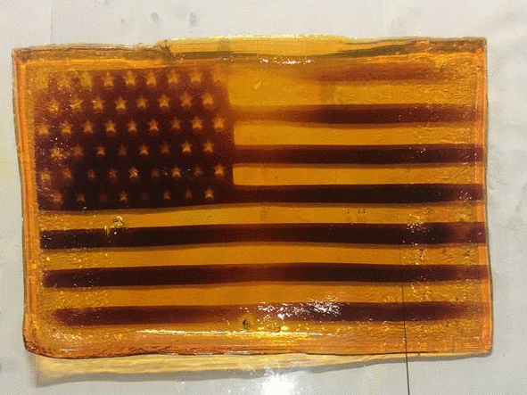
The photopattern fades from hydrogel with time and exposure to air. (Image courtesy of Barnes laboratory)
Now that researchers have spatial control over the activation of the hydrogel, they can turn to optimizing it for biomedical applications. Barnes is collaborating with Moe R. Mahjoub at the Washington University School of Medicine to that end.
Researchers will next focus on showing that the hydrogels are durable enough to support applications with cells suspended in a 3D matrix. Being able to activate specific areas in three dimensions is a key step toward successfully growing tissue in a 3D cell culture. Further refinements to the material will include activating it with other wavelengths of light, such as infrared, which would allow noninvasive activation through human tissue.
Read more on the chemistry website.


Comments and respectful dialogue are encouraged, but content will be moderated. Please, no personal attacks, obscenity or profanity, selling of commercial products, or endorsements of political candidates or positions. We reserve the right to remove any inappropriate comments. We also cannot address individual medical concerns or provide medical advice in this forum.