$6.3 million for center to develop new tracers for PET scans
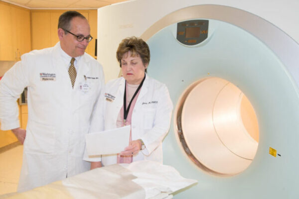
With the help of a five-year, $6.3 million NIH grant, School of Medicine radiologist Robert J. Gropler, MD, aims to help PET technology reach its potential by expanding the community of PET researchers.

Scientists use PET scans to monitor lung inflammation noninvasively
In this PET image, the arrow shows inflammation of the lungs.A noninvasive approach for assessing lung inflammation should accelerate efforts to develop drugs for inflammatory lung conditions like cystic fibrosis and pneumonia, scientists at the School of Medicine report. Researchers have used positron emission tomography (PET) scans to monitor artificially induced inflammation in the lungs of healthy volunteers. The new imaging process may help doctors monitor the conditions of patients with inflammatory lung diseases and should make it easier for investigators to test potential anti-inflammatory drugs.
Scientists use PET scans to monitor lung inflammation noninvasively
In this PET image, the arrow shows inflammation of the lungs.A noninvasive approach for assessing lung inflammation should accelerate efforts to develop drugs for inflammatory lung conditions like cystic fibrosis and pneumonia, scientists at Washington University School of Medicine in St. Louis report. Researchers have used positron emission tomography (PET) scans to monitor artificially induced inflammation in the lungs of healthy volunteers. The new imaging process may help doctors monitor the conditions of patients with inflammatory lung diseases and should make it easier for investigators to test potential anti-inflammatory drugs.
Imaging technique detects plaques in Alzheimer’s disease
PET scans: normal (top 2 rows) and Alzheimer’s.After decades of searching, scientists finally may have identified a way to study Alzheimer’s disease changes in living human brains. Researchers at Washington University’s Alzheimer’s Disease Research Center (ADRC) have begun studies of an imaging agent developed at the University of Pittsburgh that could potentially detect amyloid plaques with a positron emission tomography (PET) scan, enabling earlier detection of the disease and improved testing of new treatments.
Low receptor levels and low mood
Areas of red and yellow show increased uptake of the altanserin tracer due to binding to the serotonin receptors.Most of us feel sad from time to time, even very sad, but during a bout of clinical depression, a person is unable to escape their low mood for several weeks at a time. A popular and effective treatment for depression involves a class of drugs called selective serotonin reuptake inhibitors (SSRIs). Those drugs increase serotonin levels in the brain and help relieve symptoms of depression, and most scientists believe the brain chemical serotonin plays a key role in depression. Now neuroscience researchers at Washington University School of Medicine in St. Louis have found that in people who are depressed, a key brain structure has an abnormally low number of cellular serotonin receptors.
‘Visualizing’ Tourette Syndrome
Sophisticated brain imaging reveals that several brain regions can become overactivated when people with Tourette Syndrome perform tasks related to memory.Neuroscience researchers at Washington University School of Medicine in St. Louis are studying the brains of patients with Tourette Syndrome (TS) to see whether they can use sophisticated imaging techniques to identify differences in the dopamine system of people with the tics that characterize TS. A team of researchers, led by Kevin J. Black, M.D., assistant professor of psychiatry, neurology and radiology at Washington University School of Medicine, is using PET imaging to see what the brain does in response to levodopa, a natural amino acid that has been used for many years to treat movement disorders, such as Parkinson’s disease. With PET imaging, the researchers can measure the boost in the brain’s dopamine levels in response to the drug both in people with Tourette Syndrome and in those who do not have tics. By identifying differences, they hope to isolate the causes of tics and to help people with TS control or eliminate them.