
Washington People: Sally Schwarz
The nuclear pharmacist with a flair for design is working to keep Mallinckrodt Institute of Radiology at the forefront of research and patient care.
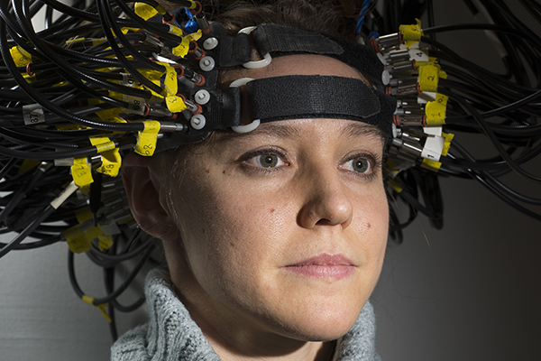
Optical brain scanner goes where other brain scanners can’t
Scientists have advanced a brain-scanning technology that tracks what the brain is doing by shining dozens of tiny LED lights on the head. The technique compares favorably to other approaches but avoids the radiation exposure and bulky magnets the others require, according to new research at the School of Medicine.
Combination PET-MRI scanner expands imaging frontiers
p, ,
{margin:0in;margin-bottom:.0001pt;font-size:12.0pt;font-family:Cambria;}
.t
{font-size:10.0pt;font-family:Cambria;}
@page WordSection1
{size:8.5in 11.0in;margin:1.0in 1.25in 1.0in 1.25in;}
div.WordSection1
{page:WordSection1;}
Scientists at Washington University School of Medicine are using a new imaging device that simultaneously performs positron-emission tomography (PET) and magnetic resonance imaging (MRI) scans, producing more detailed images than either technique alone. The scanner’s power and versatility will enable
many wonderful applications in areas ranging from cancer to neurological
disorders to heart and lung disease.
Surgery to prevent stroke causes too many complications
An operation for preventing repeat strokes in high-risk patients has failed in a multi-institutional clinical trial, led by Colin P. Derdeyn, MD, professor of radiology at Washington University School of Medicine. Results are reported in the Journal of the American Medical Association.
MRI scans reveal brain changes in people at genetic risk for Alzheimer’s
People with a known, high risk for Alzheimer’s disease develop abnormal brain function even before the appearance of telltale, amyloid plaques that are characteristic of Alzheimer’s disease, according to a new study from researchers at Washington University School of Medicine in St. Louis. The findings suggest that a gene variant affects brain function long before the brain begins accumulating the amyloid that will eventually lead to dementia.
PET scans after therapy improve cervical cancer survival predictions
GrigsbyDoctors regularly use positron emission tomography (PET) scans to diagnose cervical cancer, taking advantage of the technique’s ability to highlight metabolic differences in cancerous tissues. But PET is rarely used for follow-up assessment of cervical cancer patients after treatment. A study in the June 1 issue of Journal of Clinical Oncology shows that post-treatment PET scans could help physicians better predict which patients are largely cancer-free as a result of their treatment and which patients may soon be likely to need additional treatment.
Brain’s ‘resting’ network offers powerful new method for early Alzheimer’s diagnosis
Image courtesy of Cindy LustigParts of the brain involved in a “resting network” show large differences between young adults, older adults, and people with Alzheimer’s disease.Researchers tracking the ebb and flow of cognitive function in the human brain have discovered surprising differences in the ability of younger and older adults to shut down a brain network normally active during periods of passive daydreaming. The differences, which are especially pronounced in people with dementia, may provide a clear and powerful new method for diagnosing individuals in the very early stages of Alzheimer’s disease.
New imaging techniques help guide liver surgery
Image courtesy of William C. Chapman, M.D.William Chapman monitors his surgical instrument’s position on corresponding CT scans during liver surgery.Despite being the largest vital organ in the body, the liver has very few identifiable landmarks to help guide a cancer surgeon around its surface. Two-dimensional ultrasound images currently are the standard navigational tool, making it difficult to discern depth and location in the liver during surgery to remove tumors. That’s why a research team led by William C. Chapman, M.D., professor of surgery and chief of the Abdominal Transplantation Section at Washington University School of Medicine in St. Louis plans to launch trials examining the use of three-dimensional imaging techniques to complement ultrasound during liver surgery. The research team will investigate standard three-dimensional imaging techniques like MRI, CT scanning and PET for guiding surgeons during tumor removal surgery.
WUSTL research spotlighted at Society of Nuclear Medicine meeting, June 21-25
Smoking is more common among kids with attention deficit-hyperactivity disorder (ADHD) than those without the disorder, but the risk for smoking rises dramatically in those with the inattentive subtype of ADHD.Advances in medical imaging techniques are among the breakthroughs being presented by Washington University researchers at the Society of Nuclear Medicine’s 50th Annual Meeting June 21-25 at the Ernest N. Morial Convention Center in New Orleans, La. More than 3,600 specialists in the field of nuclear medicine are expected to attend the meeting, which focuses on current issues in nuclear medicine, including bioethics, terrorism using radioactive materials, and controversial topics in the future of PET. WUSTL-related news from the meeting includes research on a new MicroPet technique for improved imaging of small animals and a study suggesting that FDHT-PET scanning of androgen receptors (AR) is successful in imaging patients with prostate cancer. Washington University cancer imaging specialist Barry A. Siegel will receive the Society of Nuclear Medicine’s (SNM) 2003 Georg Charles de Hevesy Nuclear Pioneer Award for his distinguished contributions to nuclear medicine.