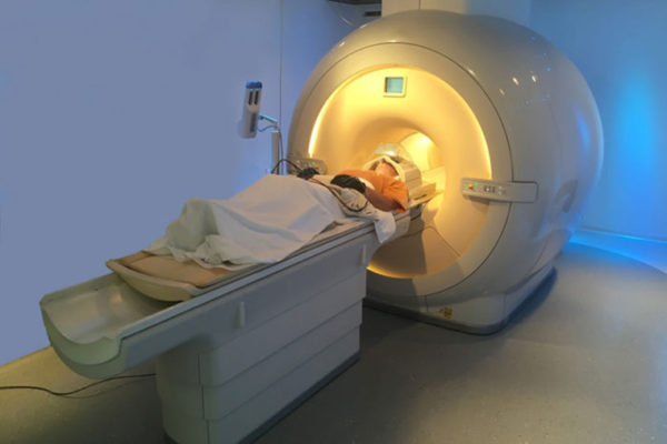
MRI scans shows promise in predicting dementia
Doctors may one day be able to gauge a patient’s risk of dementia with an MRI scan, according to a new study from the School of Medicine. Using a new technique for analyzing MRI data, researchers were able to predict who would experience cognitive decline with 89 percent accuracy.
New understanding of stroke damage may aid recovery
Stroke can lead to a wide range of problems such as depression and difficulty moving, speaking and paying attention. A new study led by Maurizio Corbetta, MD, at the School of Medicine has found evidence that stroke damage to “cables” buried inside the brain plays an important role in these impairments.

MRI for prostate biopsies increases odds of finding aggressive tumors
Prostate biopsies performed using magnetic resonance imaging are more likely to find aggressive tumors than those that rely on ultrasound, suggests a new study led by Gerald Andriole, MD, chief of urology at the School of Medicine.
Human Connectome Project releases major data set on brain connectivity
The Human Connectome Project, a five-year endeavor to
link brain connectivity to human behavior, has just released a set of
high-quality imaging and behavioral data to the scientific community. Shown is a map of the average “functional connectivity” in the human cerebral cortex, collected on healthy subjects while “at rest” in the MRI scanner.
Study to analyze brains of kids with rare disorder
School of Medicine researchers have received a five-year, $2.7 million grant to detect and analyze differences in the brains of children with a rare illness, Wolfram syndrome. The disorder includes a severe form of diabetes, hearing and vision loss and kidney problems. Patients also eventually lose muscle control and coordination from brain degeneration.
NFL funds study of the brain after concussions
Neurologists at Washington University School of Medicine in St. Louis have received funding to study the brain following repeat concussions. The project is one of 15 around the country selected by NFL Charities, the charitable foundation of the National Football League Owners.
Heavy lifting
Workers prepare to place a new MRI scanner into the East Building at the School of Medicine June 11. The 18,000-pound scanner will be used for the Human Connectome Project, a research study that will trace the anatomical and functional connections between different
regions of the brain’s gray matter.
Ultrasound screening finds more breast cancer, false positives may outweigh that benefit
Adding ultrasound exams to annual breast cancer
screening can detect more cancers in women who have
dense breasts and are at a higher risk of breast cancer, according to a
three-year, multi-center trial appearing this week in the Journal of the American Medical Association. But the scans carry risks that may outweigh their benefits.
Combination PET-MRI scanner expands imaging frontiers
p, ,
{margin:0in;margin-bottom:.0001pt;font-size:12.0pt;font-family:Cambria;}
.t
{font-size:10.0pt;font-family:Cambria;}
@page WordSection1
{size:8.5in 11.0in;margin:1.0in 1.25in 1.0in 1.25in;}
div.WordSection1
{page:WordSection1;}
Scientists at Washington University School of Medicine are using a new imaging device that simultaneously performs positron-emission tomography (PET) and magnetic resonance imaging (MRI) scans, producing more detailed images than either technique alone. The scanner’s power and versatility will enable
many wonderful applications in areas ranging from cancer to neurological
disorders to heart and lung disease.
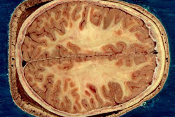
Scientists have new help finding brain’s nooks and crannies
Like explorers mapping a new planet, scientists probing the brain need every type of landmark they can get. Each mountain, river or forest helps scientists find their way through the intricacies of the human brain. Researchers at Washington University School of Medicine in St. Louis have developed a new technique that provides rapid access to brain landmarks formerly only available at autopsy.
View More Stories