Tip sheets highlight timely news and events at Washington University in St. Louis. For more information on any of the stories below or for assistance in arranging interviews, please see the contact information listed with each story.
Supersizing America
Supersized servings and bigger beverages build bulging bellies

Obesity puts people at risk for heart disease, diabetes, stroke, high blood pressure and cancer. It also decreases quality of life. But that’s not stopping Americans from eating and drinking more than ever before. Almost two-thirds of Americans are either overweight or obese, and a major factor contributing to increasing body sizes appears to be increasing portion sizes. Obesity researcher Samuel Klein, M.D., the Danforth Professor of Medicine and Nutritional Science at Washington University School of Medicine in St. Louis, says the obesity epidemic continues to get worse in spite of a great deal of research about the dangers of being overweight and increasing numbers of people who are trying to lose weight. Part of the problem is that many people tend to eat what is put in front of them, and serving sizes are larger than ever before.
Imaging Alzheimer’s disease
MRI scans distinguish brain changes in Alzheimer’s patients from changes in normal aging
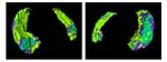
Even when people have no symptoms, their brains already may be dotted with the plaques and tangles that characterize Alzheimer’s disease. As treatments to halt the progress of Alzheimer’s disease appear on the horizon, scientists are looking for new ways to identify Alzheimer’s-associated changes in the brain before cognitive decline begins. By examining brain images, researchers, led by John G. Csernansky, M.D., the Gregory B. Couch Professor of Psychiatry, and Lei Wang, Ph.D., research associate in psychiatry, both at Washington University’s Silvio Conte Center for Neuroscience Research, found that the volume and shape of certain brain structures change in different patterns during Alzheimer’s disease than in healthy aging. They believe that someday using these imaging techniques may allow for earlier diagnosis of Alzheimer’s disease, preferably before the most devastating symptoms appear.
Countering Crohn’s Disease
Research moves forward in clinical trials
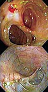
Researchers at Washington University School of Medicine in St. Louis continue to make progress in finding a potential treatment for Crohn’s disease, a chronic and serious inflammatory disease of the gastrointestinal tract that affects about half a million people in the United States. Later this month, the research team of Joshua Korzenik, M.D., assistant professor of medicine, and Brian Dieckgraefe, M.D., Ph.D., also an assistant professor of medicine in the division of gastroenterology, will present preliminary data from patients with moderate to severe Crohn’s disease who were treated at 33 centers around the United States. Patients who received daily injections of a drug called GM-CSF (granulocyte macrophage colony stimulating factor), which stimulates the activity of certain cells in the immune system, tended improve. And although the data have not yet been presented, the results from the placebo-controlled phase II treatment study were positive.
Improving survival in sepsis
How immune cells die can influence patient mortality
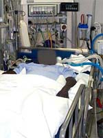
Sepsis, sometimes called blood poisoning, is the leading cause of death among critically ill patients in the United States. For many years, scientists believed it was the result of an uncontrolled inflammatory response, but several studies that involved anti-inflammatory drugs were not successful at improving survival. Now, a research team led by Richard S. Hotchkiss, M.D., professor of anesthesiology and of medicine and associate professor of surgery and of molecular biology and pharmacology, and Irene E. Karl, Ph.D., research professor of medicine at Washington University School of Medicine in St. Louis have found that how immune cells die in sepsis might be a key to whether patients survive. When immune cells die through a process known as programmed cell death, or apoptosis, a patient’s chance of survival appears to be much lower than if cells die through a different mechanism called necrosis.