New findings from researchers at Washington University School of Medicine in St. Louis suggest the eye’s cornea can resist infection from the novel coronavirus. Although the herpes simplex virus can infect the cornea and spread to other parts of the body in patients with compromised immune systems, and Zika virus has been found in tears and corneal tissue, SARS-CoV-2, the virus that causes COVID-19, does not appear to replicate in the human cornea.
The researchers have yet to determine, however, whether other tissue in and around the cornea, such as the tear ducts and the conjunctiva, are vulnerable to the virus.
The new findings are published Nov. 3 in the journal Cell Reports.
“Our findings do not prove that all corneas are resistant,” said first author Jonathan J. Miner, MD, PhD. “But every donor cornea we tested was resistant to the novel coronavirus. It’s still possible a subset of people may have corneas that support growth of the virus, but none of the corneas we studied supported growth of SARS-CoV-2.”
Miner, an assistant professor of medicine, of molecular microbiology and of pathology and immunology, teamed up with ophthalmologist Rajendra S. Apte, MD, PhD, to study mouse and human corneas exposed to the herpes simplex, Zika and SARS-CoV-2 viruses.
“Some COVID-19 patients get eye symptoms, such as conjunctivitis (pinkeye), but it’s not clear that the viral infection itself causes that; it could be related to secondary inflammation,” said Apte, the Paul A. Cibis Distinguished Professor in the John F. Hardesty Department of Ophthalmology & Visual Sciences. “The cornea and conjunctiva are known to have receptors for the novel coronavirus, but in our studies, we found that the virus did not replicate in the cornea.”
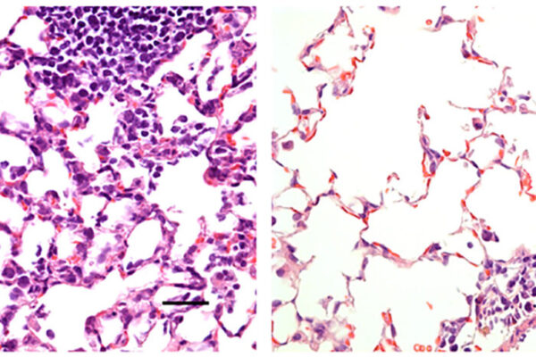
Prior research in human and mouse corneal tissue had demonstrated that Zika virus could be shed in tears, and the researchers wanted to learn whether the cornea might serve as an entry point for SARS-CoV-2. Apte, Miner and their colleagues tested that by exposing the eye tissue to the different viruses and observing whether the viruses could grow and replicate. The scientists also identified key substances in corneal tissue that can promote or inhibit viral growth.
One inhibitor they identified is called interferon lambda. They found that interferon lambda prevented efficient growth of Zika virus and herpes simplex virus in the cornea. But with SARS-CoV-2, levels of the substance had no effect on whether the virus could replicate. It simply could not gain a foothold whether interferon lambda was present or not.
That’s reassuring to Apte, also a professor of developmental biology and of medicine, who said it suggests COVID-19 probably cannot be transmitted through a cornea transplant or similar procedures in the eye.
“Our data suggest that the novel coronavirus does not seem to be able to penetrate the cornea,” Apte said.
Miner added, however, that because of unknowns involving the tear ducts and the conjunctiva, it’s too soon to dismiss the importance of eye protection.
“It’s important to respect what this virus is capable of and take appropriate precautions,” he said. “We may learn that eye coverings are not necessary to protect against infection in the general community, but our studies really are just the beginning. We need larger clinical studies to help us better understand all the potential routes of SARS-CoV-2 transmission, including the eye.”