Acute kidney injury, or acute renal failure, can occur suddenly from a variety of causes, including the systemic blood infection sepsis, which causes changes in oxygen flow to and metabolism in the kidneys. Researchers in the McKelvey School of Engineering at Washington University in St. Louis and at the University of Virginia recently developed a high-tech imaging technique that opens up opportunities to study dysfunction in acute and chronic kidney disease.

Song Hu, associate professor of biomedical engineering in the McKelvey School of Engineering, and Mark D. Okusa, the John C. Buchanan Distinguished Professor of Medicine in the Division of Nephrology and Center for Immunity, Inflammation and Regenerative Medicine at the University of Virginia Health System, led a team that used photoacoustic microscopy to image changes in oxygen delivery in the blood and oxygen metabolism in tissue in a mouse model.
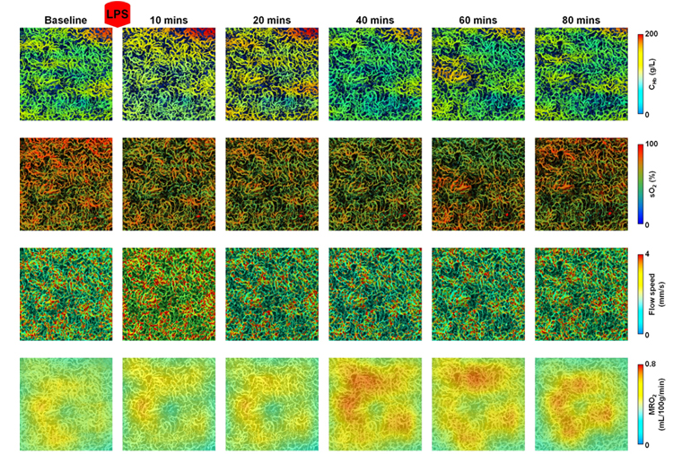
The technique, which uses a combination of light and sound to take high-resolution images at 200 microns deep, allowed the researchers to quantify the concentration of hemoglobin, oxygen saturation of hemoglobin and blood flow in tiny peritubular capillaries in the kidneys of mice with sepsis, a potentially life-threatening systemic infection.

Sepsis causes multiple changes, including inflammation and disturbed cellular metabolism, all of which lead to changes in the micro-and macrocirculatory systems, such as reduced oxygen to kidney tissues. To date, researchers have not been able to envision the mechanisms of the lack of oxygen on the kidneys due to inadequate existing imaging techniques. Hu and Okusa’s team set out to change that.
In research published in Kidney International July 2, 2021, their photoacoustic microscopy imaging technique showed that sepsis significantly reduced several biomarkers, including oxygen saturation of hemoglobin in the peritubular capillaries as well as the cellular energy levels, or ATP, in the kidney. Interestingly, early in the course following initiation of sepsis, there were minor changes in the blood flow in the capillaries and in plasma creatinine, a waste product removed from the body by the kidneys.
“Our technology provides microvascular mapping of blood oxygen in kidneys for the first time,” Hu said. “We provided a simultaneous acquisition of multiple microvascular parameters, including hemoglobin concentration, blood oxygenation and blood flow, which has been particularly difficult to get previously.”
The photoacoustic technique was able to zoom in on the peritubular capillaries, which are smaller than 10 microns in diameter, or .01 millimeters, in the mouse kidney, which is itself only about 6 to 7 millimeters in size.
Next, the team plans to use this technology to study kidney disease mechanisms in other animal models.
“While this cannot be directly applied to humans, it allows us to understand the disease mechanisms,” Hu said. “Studying the relationship between oxygen metabolic dysfunction and acute kidney injury could lead to new therapeutic targets and improved understanding of how to reverse or reduce disease damage.”
The team’s other long-term goal is to develop deep-penetrating photoacoustic microscopy techniques that would allow them to see multiple millimeters or even centimeters into the human kidney.
“That would allow us to directly apply this technology in the clinical setting,” Hu said.
In a separate project funded by the Chan-Zuckerberg Initiative, Hu is leading a group to develop a new super-resolution photoacoustic imaging technique that will enable single-cell resolution at 1 centimeter depth. That work is with Washington University’s Lan Yang, the Edwin H. & Florence G. Skinner Professor in the Preston M. Green Department of Electrical & Systems Engineering, and Adam Kepecs, BJC investigator and professor of neuroscience and psychiatry in the School of Medicine.
The McKelvey School of Engineering at Washington University in St. Louis promotes independent inquiry and education with an emphasis on scientific excellence, innovation and collaboration without boundaries. McKelvey Engineering has top-ranked research and graduate programs across departments, particularly in biomedical engineering, environmental engineering and computing, and has one of the most selective undergraduate programs in the country. With 140 full-time faculty, 1,387 undergraduate students, 1,448 graduate students and 21,000 living alumni, we are working to solve some of society’s greatest challenges; to prepare students to become leaders and innovate throughout their careers; and to be a catalyst of economic development for the St. Louis region and beyond.
This research was supported by the National Science Foundation (CBET-2023988).
Sun N, Zheng S, Rosin D, Poudel N, Yao J, Perry H, Cao R, Okusa M, Hu S. Development of a photoacoustic microscopy technique to assess peritubular capillary function and oxygen metabolism in the mouse kidney. Kidney International, July 2, 2021.DOI:https://doi.org/10.1016/j.kint.2021.06.018