Washington University School of Medicine recently teamed up with the Saint Louis Art Museum and the university’s Mildred Lane Kemper Art Museum to scan some very unusual patients: three Egyptian mummies.
The scanning took place Sunday, Oct. 12, at the Center for Advanced Medicine on the Medical Campus. The mummies, two of which are on long-term loan to the Saint Louis Art Museum from the Kemper Art Museum, were carefully transported across Forest Park and scanned one by one in a state-of-the-art computerized tomography (CT) scanner.
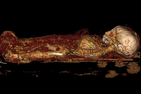
Among the early findings: One of the mummies already was known to have a brain, but scans revealed she also still has lungs. In many mummies, lungs typically were removed prior to burial.
The scientists — radiologists with the university’s Mallinckrodt Institute of Radiology — discovered that the same mummy also has an array of small objects around her head. It appears to be a headdress or embellished shroud, but other possibilities include packing material or debris.
The scientists were surprised to find that a second mummy appeared to be significantly shorter than his sarcophagus. Further scanning revealed that his head had been dislodged from his body, perhaps when grave robbers ransacked his tomb. They found an item on his chest that may have been a burial amulet missed by grave robbers. They hope to use the scanning data to reconstruct the item with a 3-D printer.
The researchers expect to have more detailed results ready to share in December.
Lisa Çakmak, PhD, assistant curator for ancient art at the Saint Louis Art Museum, initiated the project. The museum is preparing for an upcoming reinstallation of the mummies, and Çakmak thought scans might provide valuable information about the mummies and their societies that could be incorporated into the new exhibit.
The mummies’ burial containers and wrappings identify each by name. The Saint Louis Art Museum’s mummy is Amen-Nestawy-Nakht, a male; the Kemper Art Museum mummies are Pet-Menekh, also a male, and Henut-Wedjebu, a female.
Karen K. Butler, PhD, associate curator of the Kemper Art Museum, said Pet-Menekh and Henut-Wedjebu were donated to the university in 1896 by Charles Parsons, a St. Louis banker and prominent art collector. Working with a curator from the Egyptian Museum in Cairo, Parsons acquired them shortly after excavation through the Antiquities Service of Egypt.
“The mummies have been part of Washington University for more than 100 years,” Butler said. “Faculty from anthropology, classics, art history and archaeology all take students to see them. They are very much part of university life.”
Mummy history
Henut-Wedjebu, the oldest of the mummies, was discovered in a cave-like tomb near the ruins of the Egyptian city of Thebes. Her name means “singer of Amun and lady of the house,” and her elaborately gilded coffin, decorated with texts from the Book of the Dead, is one of only eight such objects to survive from the reign of Amenhotep III (1390-1353 B.C.).
Pet-Menekh — or “he whom the excellent one has given” — is thought to have been a priest of the god Chem during the Ptolemaic period (c. 300 B.C.). He died in his 30s or 40s, possibly of sudden trauma or acute disease. His coffin — likely found at the Necropolis of El-Hawawish in Akhmim — is richly decorated with hundreds of hieroglyphics as well as images of the goddesses Isis and Nut.
Amen-Nakht — or “Amun (Lord) of the Thrones of the Two Lands is Strong” — acquired by the Saint Louis Art Museum in 1980, was a priest of Amun during the 22nd Dynasty (945–712 B.C.). His coffin is thought to have been discovered in the Necropolis of Thebes. A painted cartonnage — a kind of funerary case made of linen and plaster — covers the body and illustrates the panoply of deities charged with escorting him into the afterlife.

“The technical sophistication of all three mummies suggests that these were well-off individuals,” said Çakmak. “We would expect to see that reflected in the condition of their teeth and skeletons. The CT scan helps us to better understand their lifestyles.”
Added Butler: “As a university museum, we’re always looking for new ways to understand and research our collection. This collaboration has allowed us to bring together two different aspects of the university community: art historical scholarship and scientific research. It’s really thrilling.”
Mummies present art experts and scientists with a formidable challenge: They are incredible time capsules from human societies that vanished thousands of years ago, but opening the capsules would desecrate human remains and possibly destroy unique cultural treasures.
Modern medical imaging techniques offer ways to peer into these time capsules without physically opening them. Scientists scanned Amen-Nestaway-Nakht two decades ago, but imaging technology has advanced significantly since then.
Çakmak, Butler and others approached Gil Jost, MD, then director of the Mallinckrodt Institute, about the possibility of getting the mummies scanned. Jost enlisted Sanjeev Bhalla, MD, professor of radiology and chief of cardiothoracic imaging, to lead the research team.
Other investigators for the project included Pamela Woodard, MD, professor of radiology and director of the Center for Clinical Imaging Research; Vincent Mellnick, MD, assistant professor of radiology; and Michelle Miller-Thomas, MD, assistant professor of radiology. The team represented expertise in imaging of the brain, abdomen and coronary arteries.
“This has been Dr. Jost’s specialty: bringing together the best technological resources and expertise necessary to do fascinating research, and then stepping away and letting them go to work,” said Bhalla. “His attitude always is, ‘Let’s make it happen.’”
The researchers and other medical center staff volunteered their time, and the School of Medicine and Barnes-Jewish Hospital donated time on the scanner and the computing resources necessary to process the results. The Saint Louis Art Museum paid for transporting the mummies.
How best to scan a mummy
The scientists considered scanning the mummies with a magnetic resonance imaging (MRI) unit, but it was impossible to guarantee that the mummies were free of any metals. Metal is prohibited in MRIs because the strong magnets in the scanners can damage the equipment and the subject being scanned. More importantly, though, mummies are free of water as a result of the mummification process, and the images created with MRI scanning are dependent on the water content of tissue.
The researchers instead brought the mummies to a powerful and recently installed computerized tomography (CT) scanner. The unit uses X-rays to virtually slice a solid object, producing detailed 3-D images of its interior.
“This new CT scanner has higher spatial resolution and quickly can assemble slices in a variety of ways, providing more medical details about the mummies,” Bhalla said.
In living patients, Bhalla and his colleagues often inject contrast agents that help make different types of cells and tissues stand out. This was not an option for the mummies, but researchers scanned them at two different energy levels to enhance details.
Among other goals, the researchers are analyzing the data for signs of artery hardening in the mummies. Indicators of heart disease have been detected in prior mummy scans, but it’s not clear yet if this is reflective of the elite lifestyle of anyone rich enough to be mummified or if heart disease was a common problem in ancient Egyptian society.
The researchers also will take a close look at the mummies’ teeth. The degree of wear on the teeth helps scientists more precisely estimate a mummy’s age at the time of death. They also will search for evidence of what caused the mummies’ deaths.
Logistically, the scans were complicated. The mummies had to be carefully removed from their display cases, packed and prepared for transport in custom-built boxes. The team took precise measurements to be sure each mummy would it into the scanner.
But Bhalla viewed another aspect of the scans as the greatest challenge of the project.
“It was very important for us to remember that these were human beings we were scanning,” he said. “We had to do the scanning in an atmosphere of spiritual and physical respect, and with the help of museum staff who acted as a kind of surrogate family for the mummies, we did that.”
Added Çakmak: “Mummification was a difficult and expensive process. It’s really very poignant. Each of these people was beloved by someone.”