A study in Nature Cardiovascular Research by researchers at Washington University School of Medicine in St. Louis explores a new, noninvasive imaging technique that helps scientists visualize immune cells in the human heart.
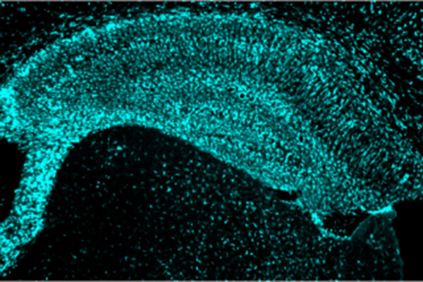
Inflammatory immune cells contribute to cardiovascular disease, increasing the risk of a heart attack and heart failure. However, technologies to identify the immune cell types responsible for inflammation in the muscular tissue of the heart — the myocardium — are limited. The researchers developed a radiotracer to help visualize a subset of inflammatory immune cells marked by the expression of C-C chemokine receptor 2, which includes harmful populations of monocytes and macrophages.
The research was performed by Yongjian Liu, PhD, a professor of radiology at Mallinckrodt Institute of Radiology (MIR), and Kory J. Lavine, MD, PhD, an associate professor of medicine, of developmental biology, and of pathology and immunology, in collaboration with colleagues from the PET Radiotracer Translation and Resource Center and Immuno-Fib HF Network.
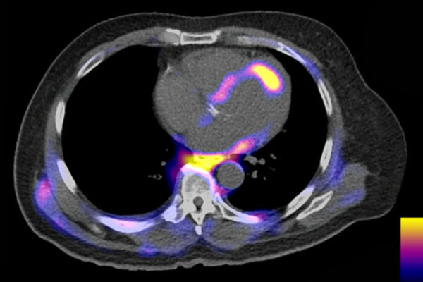
The research team established the feasibility of noninvasively visualizing inflammatory immune cells using positron emission tomography in patients who had suffered heart attacks. Their findings may help identify individuals who may benefit from immunomodulatory therapies.
Read more on the MIR website.