Biomedical engineer Abhinav Jha, an assistant professor at the McKelvey School of Engineering and of radiology at the School of Medicine, both at Washington University in St. Louis, has long advocated that artificial intelligence (AI) tools used in medical applications for image processing need to be evaluated based on clinical tasks, not visual appeal.
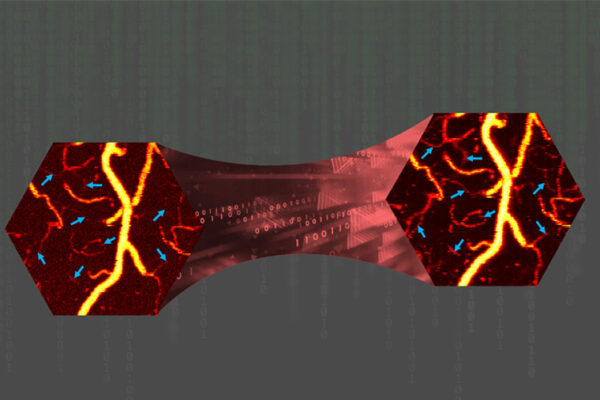
In a study published in IEEE Transactions on Radiation and Plasma Medical Sciences, Jha and his collaborators developed a tool that demonstrates the potential to improve performance on clinical tasks. The tool has been developed in the context of denoising myocardial perfusion imaging (MPI) single-photon emission computed tomography (SPECT) images.
For doctors to obtain these images, which help evaluate blood flow to the heart muscle, patients first receive a dose of radioactive tracer and then remain stationary for up to 15 minutes during the scan. Reducing the dose of the tracer, the time required or both would benefit patients, streamline the process and reduce imaging cost. However, it also would reduce the ability to visualize cardiac defects with the images.
Inspired by our understanding of how the human visual system works, Jha’s team developed a detection task-specific deep-learning-based approach for denoising these low-count MPI SPECT images so the quality improves. The new tool, called DEMIST, leverages a deep-learning framework to selectively clean such images while preserving features that influence detection tasks.
Read more on the McKelvey School of Engineering website.
Comments and respectful dialogue are encouraged, but content will be moderated. Please, no personal attacks, obscenity or profanity, selling of commercial products, or endorsements of political candidates or positions. We reserve the right to remove any inappropriate comments. We also cannot address individual medical concerns or provide medical advice in this forum.